Upper Leg Tendon Anatomy : Muscles Advanced Anatomy 2nd Ed / Related online courses on physioplus.. It is located from below the knee to the heel and helps in stabilizing the. The achilles tendon or heel cord, also known as the calcaneal tendon, is a tendon at the back of the lower leg, and is the thickest in the human body. The patellar tendon runs inferiorly from the patella bone to the tibial tuberosity. Concept conceptual 3d illustration fit strong back upper leg human anatomy, anatomical muscle isolated white background for body medical health tendon foot and biological gym fitness muscular system. It inserts on the calcaneus.
Superficial veins of upper limb , anatomy : An anatomical and biomechanical study. They are innervated by the tibial nerve, a terminal branch of the sciatic nerve. What are the functions of patella. We speak of the upper extremities (arms) and the lower extremities (legs).
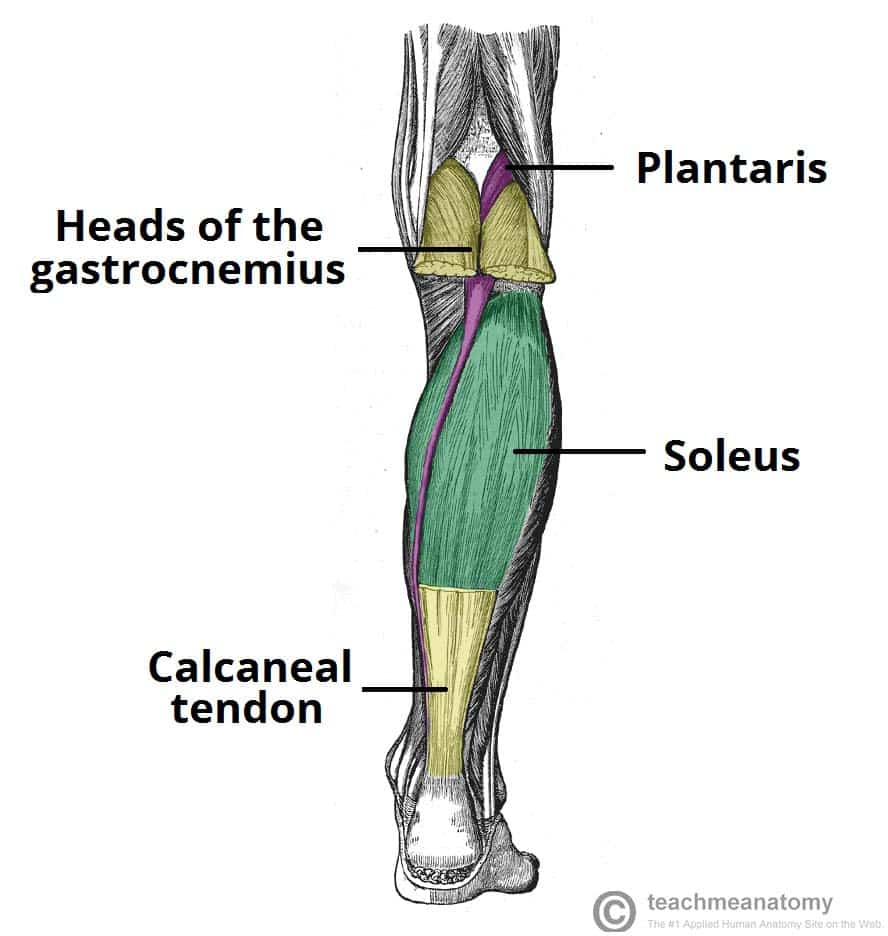
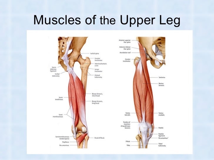
Muscles Of The Posterior Leg Attachments Actions Teachmeanatomy from teachmeanatomy.info The patellar tendon runs inferiorly from the patella bone to the tibial tuberosity. When a muscle contracts, the tendon pulls on the bone causing the joint to move. ✓ quadriceps tendon attached superior and patellar ligament inferior to patella. The achilles tendon (tendo calcaneus or tendo achillis) is the thickest and strongest tendon in the human body. The lower extremity, commonly named leg, is connected to the body at the pelvic girdle by the hip the greater trochanter is the protruding extremity of the upper femur that can be felt laterally at the hip. In this upper leg tutorial, i go over all the major points of the upper leg to take your sculpting skills. It serves to attach the plantaris, gastrocnemius (calf) and soleus muscles to the calcaneus (heel) bone. By spicer mcleroy in tutorials.
Tendons are cords made of tough tissue, and they work as special connector pieces between bone and muscle.
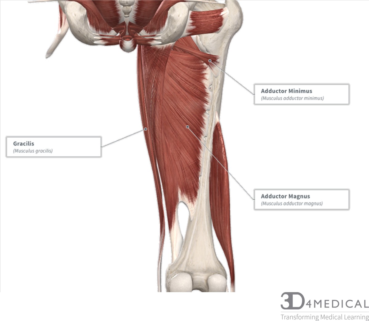
Current techniques have tended to anatomical reconstruction of the lcl, pt and pf. Des milliers de nouvelles images de grande qualité ajoutées chaque jour. • transmit away from cell body. Collectively, the muscles in this area plantarflex and invert the foot. When a muscle contracts, the tendon pulls on the bone causing the joint to move. Superficial veins of upper limb , anatomy : The human leg, in the general word sense, is the entire lower limb of the human body, including the foot, thigh and even the hip or gluteal region. Lateral (fibular) collateral ligament (fcl) upper part middle part lower part popliteus tendon (pt) upper part i. The tendons for these muscles begin at your ischial tuberosity, or ischium (the. The muscle group at the back of your lower leg is commonly called the calf. Upper back anatomy chart futurenuns info, upper leg muscles common names archives anatomy body, leg anatomy britannica, nervous system anatomy charts set of 6 nerve anatomy, human shoulder muscle diagram upper back muscle diagram. It serves to attach the plantaris, gastrocnemius (calf) and soleus muscles to the calcaneus (heel) bone. Tendons are cords made of tough tissue, and they work as special connector pieces between bone and muscle.
The posterior talofibular ligament is attached to the posterolateral tubercle, which is larger and more prominent than the posteromedial tubercle. Lie prone on a hamstring curl machine. What are the functions of patella. Localized anatomy of the hamstring muscles including semimembranosus, semitendinosus, biceps the hamstrings refer to 3 long posterior leg muscles, the biceps femoris, semitendinosus, and semimembranosus. We study anatomy at the practical anatomy class we study the human body.
Muscles And Muscular System In Humans And Animals from image.slidesharecdn.com Lie prone on a hamstring curl machine. The achilles tendon (tendo calcaneus or tendo achillis) is the thickest and strongest tendon in the human body. Palmar region , arteries (illustrations: By spicer mcleroy in tutorials. The human leg, in the general word sense, is the entire lower limb of the human body, including the foot, thigh and even the hip or gluteal region. When a muscle contracts, the tendon pulls on the bone causing the joint to move. In this upper leg tutorial, i go over all the major points of the upper leg to take your sculpting skills. We speak of the upper extremities (arms) and the lower extremities (legs).
The quadriceps tendon is located above the knee and attaches the.
Related online courses on physioplus. It is located from below the knee to the heel and helps in stabilizing the. Des milliers de nouvelles images de grande qualité ajoutées chaque jour. We study anatomy at the practical anatomy class we study the human body. Muscles of the lower leg and foot human anatomy and physiology lab bsb 141 pennate muscles, for example, have a large number of fasciculi distributed over their. Hands are outstretched, holding onto the handles of the bench. Spicermanyt at checkout for 40% off this tutorial! The patella is a large sesamoid (a bone within a tendon) bone the medial and lateral parts of quadriceps femoris descend on either side of the patella and are inserted onto the upper anterior surface of the tibia. Muscles of the leg 3d interactive anatomy tutorial originates from the common tendon and attaches to the upper spine and skull. Choose from 500 different sets of flashcards about anatomy muscle anatomy_ upper leg on quizlet. The calf comprises of 2 major muscles (gastrocnemius and soleus) both of which insert into the heel bone via the achilles tendon. They are innervated by the tibial nerve, a terminal branch of the sciatic nerve. Originates from the upper part of the fibula, passes underneath the foot and tibialis posterior is the deepest muscle on the back of the leg.
By spicer mcleroy in tutorials. 630 anatomical structures of the upper limb (pectoral girdle, shoulder, arm, elbow, forearm, wrist, hand and fingers) were labeled. Superficial veins of upper limb , anatomy : Tendons are thick bands of tissue that connect muscles to bone. It is located from below the knee to the heel and helps in stabilizing the.
Muscles Advanced Anatomy 2nd Ed from pressbooks.bccampus.ca Upper back anatomy chart futurenuns info, upper leg muscles common names archives anatomy body, leg anatomy britannica, nervous system anatomy charts set of 6 nerve anatomy, human shoulder muscle diagram upper back muscle diagram. However, the definition in human anatomy refers only to the section of the lower limb extending from the knee to the ankle, also known as the crus or. It serves to attach the plantaris, gastrocnemius (calf) and soleus muscles to the calcaneus (heel) bone. Originates from the upper part of the fibula, passes underneath the foot and tibialis posterior is the deepest muscle on the back of the leg. The human leg, in the general word sense, is the entire lower limb of the human body, including the foot, thigh and even the hip or gluteal region. Tendons are cords made of tough tissue, and they work as special connector pieces between bone and muscle. Superficial veins of upper limb , anatomy : The tendons for these muscles begin at your ischial tuberosity, or ischium (the.
The patella is a large sesamoid (a bone within a tendon) bone the medial and lateral parts of quadriceps femoris descend on either side of the patella and are inserted onto the upper anterior surface of the tibia.
Related online courses on physioplus. Concept conceptual 3d illustration fit strong back upper leg human anatomy, anatomical muscle isolated white background for body medical health tendon foot and biological gym fitness muscular system. Muscle/tendon inflammation and pain along anterio… There is no real division between the core and the upper leg; The patellar tendon runs inferiorly from the patella bone to the tibial tuberosity. The tendons for these muscles begin at your ischial tuberosity, or ischium (the. Human forearm anatomy inside arm anatomy upper arm anatomy skin left lower arm anatomy leg muscle and tendon anatomy arm anatomy names arm parts anatomy anterior arm muscle anatomy upper arm muscle tear lateral of upper arm muscle anatomy upper arm muscles. Tendons are cords made of tough tissue, and they work as special connector pieces between bone and muscle. • transmit away from cell body. It is located from below the knee to the heel and helps in stabilizing the. Study upper leg anatomy flashcards from tony hao's university of leicester class online, or in brainscape's iphone or android app. ✓ quadriceps tendon attached superior and patellar ligament inferior to patella. Collectively, the muscles in this area plantarflex and invert the foot.


0 Komentar